Pages
▼
Sunday, 31 March 2013
HOLT ORAM SYNDROME
HOLT ORAM SYNDROME
BACKGROUND: Also called as heart-hand syndrome, is an inherited disorder, first described in 1960 by Holt and Oram in a 4 generation family with ASD and thumb abnormalities
GENETICS: Autosomal dominant inheritance with complete penetrance. Due to mutations in TBX5 on long arm of chromosome 12, which is important in development of heart and hand
Clinical features:
Hand defects: Always present. Includes carpal bone fusion, absent radius, absent or triphalangeal thumb
Cardiac defects: Present in 75% of patients. Includes ASD(mc), VSD, conduction defects, AF, anamolous venous return
PROGNOSIS: Depends on severity of cardiac lesions. In mild lesions near normal life expectancy.
This is an image from a Holt Oram syndrome patient showing hand abnormalities
Clinical Features in Patients with Wilson Disease
Clinical Features in Patients with Wilson Disease
Hepatic
· Asymptomatic hepatomegaly
· Isolated splenomegaly
· Persistently elevated serum aminotransferase activity (AST, ALT)
· Fatty liver
· Acute hepatitis
· Resembling autoimmune hepatitis
· Cirrhosis: compensated or decompensated
· Acute liver failure
Neurological
· Movement disorders (tremor, involuntary movements)
· Drooling, dysarthria
· Rigid dystonia
· Pseudobulbar palsy
· Dysautonomia
· Migraine headaches
· Insomnia
· Seizures
Psychiatric
· Depression
· Neurotic behaviours
· Personality changes
· Psychosis
Other Systems
· Ocular: Kayser-Fleischer rings, sunflower cataracts
· Cutaneous: lunulae ceruleae
· Renal abnormalities: aminoaciduria and nephrolithiasis
· Skeletal abnormalities: premature osteoporosis and arthritis
· Cardiomyopathy, dysrhythmias
· Pancreatitis
· Hypoparathyroidism
· Menstrual irregularities; infertility, repeated miscarriages
Image Courtesy: www.eurowilson.org
Text Reference: http://www.wilsonsdisease.org
Thursday, 28 March 2013
Human chorionic gonadotropin
Info: Few points regarding
Info: Few points regarding Human chorionic gonadotropin
• hCG can be detected in maternal urine after 7-8days of ovulation
• The rapidly rising hCG seen between 3-4 and 9-10 weeks gestation coincides with the proliferation of immature trophoblastic villi and the extent of the syncytial layer
• Plasma levels increase, doubling in concentration every 2-3 days between 60 and 90 days of gestation.
• The average peak hCG level is approximately 110,000 mIU/mL and occurs at 10 weeks gestation
• Between 12 and 16 weeks, average hCG decreases rapidly with the concentration
•Levels continue to fall from 16 to 22 weeks at a slower rate.
•During the third trimester mean hCG levels rise in gradual, yet significant, manner from 22 weeks until term.
•hCG levels are comparatively higher in women bearing female fetuses.
•Human chorionic gonadotropin may stimulate steroidogenesis in the early fetal testes resulting in virilization and sexual differentiation in males
Text Source: endotext.org
Image: acubaby.com
Saturday, 23 March 2013
Diagnosis of alcohol dependence: CAGE questionnaire
A clinically helpful approach to diagnosis of alcohol dependence and abuse is the use of the CAGE questionnaire, which is recommended in all medical history-taking.
One "yes" response should raise suspicion of an alcohol use problem, and more than one is a strong indication that abuse or dependence exists.
One "yes" response should raise suspicion of an alcohol use problem, and more than one is a strong indication that abuse or dependence exists.
Friday, 22 March 2013
Types of encephalopathy.
Some examples include:
•Mitochondrial encephalopathy: Metabolic disorder caused by dysfunction of mitochondrial DNA. Can affect many body systems, particularly the brain and nervous system.
•Glycine encephalopathy: A genetic metabolic disorder involving excess production of glycine
•Hepatic encephalopathy: Arising from advanced cirrhosis of the liver
•Hypoxic ischemic encephalopathy: Permanent or transitory encephalopathy arising from severely reduced oxygen delivery to the brain
•Static encephalopathy: Unchanging, or permanent, brain damage
•Uremic encephalopathy: Arising from high levels of toxins normally cleared by the kidneys—rare where dialysis is readily available
•Wernicke's encephalopathy: Arising from thiamine deficiency, usually in the setting of alcoholism
•Hashimoto's encephalopathy: Arising from an auto-immune disorder
•Hypertensive encephalopathy: Arising from acutely increased blood pressure
•Chronic traumatic encephalopathy: Progressive degenerative disease associated with multiple concussions and other forms of head injury
•Lyme encephalopathy: Arising from Lyme disease bacteria, including Borrelia burgdorferi.
•Toxic encephalopathy: A form of encephalopathy caused by chemicals, often resulting in permanent brain damage
•Toxic-Metabolic encephalopathy: A catch-all for brain dysfunction caused by infection, organ failure, or intoxication
•Transmissible spongiform encephalopathy: A collection of diseases all caused by prions, and characterized by "spongy" brain tissue (riddled with holes), impaired locomotion or coordination, and a 100% mortality rate. Includes bovine spongiform encephalopathy (mad cow disease), scrapie, and kuru among others.
•Neonatal encephalopathy: An obstetric form, often occurring due to lack of oxygen in bloodflow to brain-tissue of the fetus during labour or delivery
•Salmonella encephalopathy : A form of encephalopathy caused by food poisoning (especially out of peanuts and rotten meat) often resulting in permanent brain damage and nervous system disorders.
•Encephalomyopathy: A combination of encephalopathy and myopathy. Causes may include mitochondrial disease (particularly MELAS) or chronic hypophosphatemia, as may occur in cystinosis.
Thursday, 21 March 2013
Tuesday, 19 March 2013
High heels are dangerous
"According to the American Orthopaedic Foot and Ankle Society, people take an average of 10,000 steps a day. High heels shift the force of each of those steps so that the most pressure ends up on the ball of the foot and on the bones at the base of the toes. (If you wear flats, the entire foot would absorb this impact.) A 3-inch heel -- most experts consider a heel "high" at 2 inches or more -- creates three to six times more stress on the front of the foot than a shoe with a modest one-inch heel.
As a result, heels can lead to bunions, heel pain, toe deformities, shortened Achilles tendons, and trapped nerves. In fact, women account for about 90% of the nearly 800,000 operations each year for bunions, hammertoes (a permanent deformity of the toe joint in which the toe bends up slightly and then curls downward, resting on its tip), and trapped nerves, and most of these surgeries can be linked back to their high-heeled shoe choice.
The problems can travel upward, too. The ankle, knee, and hip joints can all suffer from your footwear preferences. When you walk in flats, the muscles of the leg and thigh have an opportunity to contract as well as to stretch out. However, when wearing your high-heeled shoes, the foot is held in a downward position as you walk. This keeps the knee, hip, and low back in a somewhat flexed position, which prevents the muscles that cross the backside of these joints to stretch out as they normally would. Over time, this can lead to stiffness, pain, and injury. High heels can also cause lower back strain, because the heel causes your body to pitch forward more than normal, putting excess pressure on the back."
Monday, 18 March 2013
Chest X-ray: Transposition of the Great Arteries
Chest X-ray: Transposition of the Great Arteries
The aorta and pulmonary artery are transposed so that the aorta arises from the right ventricle and the pulmonary artery arises from the left ventricle.
Chest X-ray may appear normal in newborns with transposition of the great arteries (TGA) and intact ventricular septum but may demonstrate the classic 'egg on side' appearance (heart is slightly enlarged and appears like an egg lying on its side, narrow vascular pedicle because aorta and pulmonary artery lie one in front of the other, and increased vascular lung markings).
Sunday, 17 March 2013
INTRAVENOUS CANNULAS, COLOURS ,GAUGE and FLOW RATE
INTRAVENOUS CANNULAS, COLOURS ,GAUGE and FLOW RATE::
1. ORANGE - Gauge 14 - Flow 200 ml/min
2. GREY - Gauge 16 - Flow 140 ml/min
3. GREEN - Gauge 18 - Flow 90 ml/min
4. PINK - Gauge 20 - Flow 61 ml/min
5. BLUE - Gauge 22 - Flow 36 ml/min
6. YELLOW - Gauge 24 - Flow 20 ml/min
1. ORANGE - Gauge 14 - Flow 200 ml/min
2. GREY - Gauge 16 - Flow 140 ml/min
3. GREEN - Gauge 18 - Flow 90 ml/min
4. PINK - Gauge 20 - Flow 61 ml/min
5. BLUE - Gauge 22 - Flow 36 ml/min
6. YELLOW - Gauge 24 - Flow 20 ml/min
X-ray findings of emphysema
What are the x-ray findings of emphysema?
- Lungs are large and hyperinflated.
- Signs of hyperinflation are:
- Low set diaphragm
- Flat diaphragm best determined by lateral chest
- Hyperlucent lung fields
- Increased AP diameter
- Increased retrosternal air
- Vertical heart
- Signs of hyperinflation can be seen in emphysema, chronic bronchitis and asthma.
- We can call it emphysema only when hyperinflation is associated with blebs and paucity of vascular markings in the outer third of the film.
- Lateral chest is best to evaluate flattening of diaphragm, AP diameter and retrosternal air.
This lateral chest shows:
- Increased AP diameter
- Low set flat diaphragms
- Hyperlucent lung fields
- Increased retrosternal air encroaching on heart density
- Multiple blebs: Avascular zones surrounded by thin wall
Chest X-ray: The bulging fissure sign
Chest X-ray: The bulging fissure sign
Klebsiella Pneumonia is much more common in alcoholics, elderly patients and those with long standing DM. Typically it causes a "currant-jelly sputum". On X-rays, the bulging fissure sign is specifically associated with Klebsiella infection.
Klebsiella Pneumonia is much more common in alcoholics, elderly patients and those with long standing DM. Typically it causes a "currant-jelly sputum". On X-rays, the bulging fissure sign is specifically associated with Klebsiella infection.
CERVICAL RIBS: X-ray
Xray :CERVICAL RIBS
A cervical rib is an extra rib that forms above the normal first rib, growing from the base of the neck just above the collarbone. The defect is present at birth, but usually not noticed until later in life.
Symptoms::
If the extra rib does press on a vessel or nerve, you may have any of the following symptoms:
pain in the shoulder and neck, which spreads into the arm: this may come and go or be constant
moments where you lose feeling and have weakness or
tingling in the affected arm and fingers
moments where you can't carry out fine hand movements, such as doing up buttons
Raynaud's phenomenon, where the blood vessels go into a temporary spasm, affecting blood supply to the fingers and toes (turning them white)
a blood clot forming in the subclavian artery, which can affect the blood supply to the fingers, causing small patches of red or black discolouration
swelling in the affected arm (although this is rare)
Chest X-ray: Coarctation of Aorta: Figure 3 sign
Chest X-ray: Coarctation of Aorta: Figure 3 sign
Post-stenotic dilation of the aorta results in a classic 'figure 3 sign' on x-ray. The characteristic bulging of the sign is caused by dilatation of the aorta due to an indrawing of the aortic wall at the site of cervical rib obstruction, with consequent post-stenotic dilation. This physiology results in the '3' image for which the sign is named
Post-stenotic dilation of the aorta results in a classic 'figure 3 sign' on x-ray. The characteristic bulging of the sign is caused by dilatation of the aorta due to an indrawing of the aortic wall at the site of cervical rib obstruction, with consequent post-stenotic dilation. This physiology results in the '3' image for which the sign is named
Chest X-ray: Cardiac tamponade
Chest X-ray: Cardiac tamponade
Cardiac tamponade secondary to tuberculosis in a 32-year-old man with acquired immunodeficiency syndrome.
Chest radiograph shows significant enlargement of the cardiac silhouette with the characteristic “water bottle” appearance.
Cardiac tamponade secondary to tuberculosis in a 32-year-old man with acquired immunodeficiency syndrome.
Chest radiograph shows significant enlargement of the cardiac silhouette with the characteristic “water bottle” appearance.
Chest X-Ray: Total anomalous pulmonary venous connection
Chest X-Ray: Total anomalous pulmonary venous connection
Snowman sign or `figure of 8 configuration` on chest radiograph
Snowman sign or `figure of 8 configuration` on chest radiograph
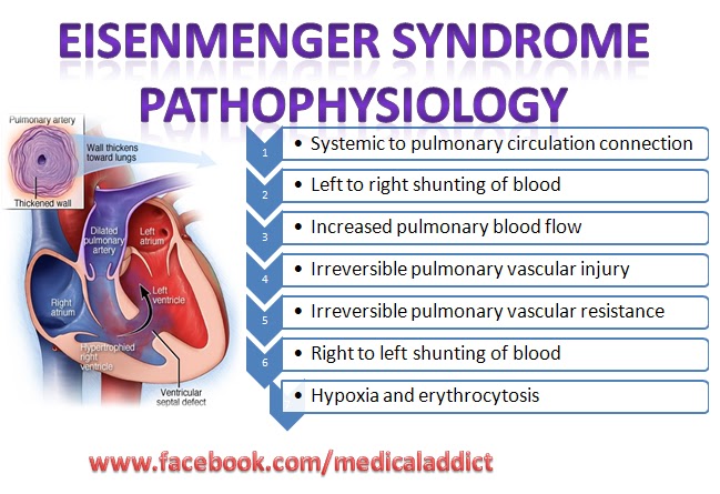
Eisenmenger syndrome Pathophysiology
Eisenmenger syndrome:
Eisenmenger's syndrome (or Eisenmenger's reaction or tardive cyanosis) is defined as the process in which a left-to-right shunt caused by a congenital heart defect causes increased flow through the pulmonary vasculature, causing pulmonary hypertension,which in turn causes increased pressures in the right side of the heart and reversal of the shunt into a right-to-left shunt.
Eisenmenger's syndrome (or Eisenmenger's reaction or tardive cyanosis) is defined as the process in which a left-to-right shunt caused by a congenital heart defect causes increased flow through the pulmonary vasculature, causing pulmonary hypertension,which in turn causes increased pressures in the right side of the heart and reversal of the shunt into a right-to-left shunt.
Chest X-ray: Nocardial pneumonia
Chest X-ray: Nocardial pneumonia.
The most common form of nocardial disease in the respiratory tract.
A dense infiltrate with a possible cavity and several nodules are apparent in the right lung.
Sulfonamides are the drugs of choice
The most common form of nocardial disease in the respiratory tract.
A dense infiltrate with a possible cavity and several nodules are apparent in the right lung.
Sulfonamides are the drugs of choice
HOW TO SURVIVE A HEART ATTACK WHEN YOU ARE ALONE?
HOW TO SURVIVE A HEART ATTACK WHEN YOU ARE ALONE??
Since many people are alone when they suffer a heart attack, without help,the person whose heart is beating improperly and who begins to feel faint, has only about 10 seconds left before losing consciousness.
However,these victims can help themselves by coughing repeatedly and very vigorously. A deep breath should be taken before each cough, and the cough must be deep and prolonged, as when producing sputum from deep inside the chest.
A breath and a cough must be repeated about every two seconds without let-up until help arrives, or until the heart is felt to be beating normally again.
Deep breaths get oxygen into the lungs and coughing movements squeeze the heart and keep the blood circulating. The squeezing pressure on the heart also helps it regain normal rhythm. In this way, heart attack victims can get to a hospital. Tell as many other people as possible about this. It could save their lives!!
A cardiologist says If everyone who sees this post shares it to 10 people, you can bet that we'll save at least one life..
Rather than sharing jokes only please contribute by forwarding this info which can save a person's life..
‘Snowman’ or ‘cottage loaf’ heart shape due to total anomalous pulmonary venous drainage
This is a classic picture of the ‘snowman’ or ‘cottage loaf’ heart shape due to total anomalous pulmonary venous drainage into a left superior vena cava (SVC). The confluence of pulmonary veins joins behind the heart and enters the ascending limb of the left SVC which appears as a dilated vessel on the right of the upper mediastinal edge. This large venous flow enters the transverse crossing vein or innominate vein making the convex roof of the cottage loaf, and joins the right SVC causing dilatation of the left side of mediastinum. As one looks at this film, there is, beneath these venous shadows, the lower part of the cottage loaf made up of dilated right atrium and ventricular mass which is the right ventricle. There is considerable increase in the size of pulmonary vessel arteries and veins (pulmonary plethora). This is due to increased pulmonary blood flow shown throughout the lungs.
This is from a young adult and it is unusual to find this in an adult. The picture is pathogmonic. The patient presents with signs of large atrial septal defect with mild cyanosis from the obligatory right to left shunt at atrial level.
This is from a young adult and it is unusual to find this in an adult. The picture is pathogmonic. The patient presents with signs of large atrial septal defect with mild cyanosis from the obligatory right to left shunt at atrial level.


